Peripheral Arterial Imaging
What is Peripheral Arterial Imaging?

Peripheral arterial imaging is a test that provides your doctor with information about the condition of the arteries outside of your heart. During the test, sound waves are “bounced” off the artery and a computer reconstructs the sound waves into a picture of the artery. Testing is done when your doctor suspects that your symptoms might be caused by a buildup of plaque in the arteries. This buildup can cause a narrowing or blockage of the artery. Testing may also be used to follow your progress if you have known vascular disease or have had surgery to open up blockages.
What happens during Arterial Imaging?
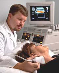
In most cases you will be asked to change into a gown. The sonographer (technician) will place some gel on your skin and then use a smooth probe to gather images of your blood vessels. Testing takes about 30 minutes. In some situations the sonographer may need to press very firmly, however, the test should not be painful or uncomfortable.
How do I prepare for Arterial Imaging?
If you are having an abdominal or renal ultrasound you need to fast for 6 hours prior to the test. You can take medications with a small sip of water. You should wear two piece clothing. If you are having a carotid ultrasound, it is helpful if you remove heavy or cumbersome neck jewelry prior to coming for the test.
What happens after the Test?
After the test, you can go home and resume your normal activities. Your doctor will review your test and discuss the results with you at your next visit or his nurse will call you with the results.
What are the Risks and Limitations of Arterial Imaging?
There is no risk associated with arterial testing.