Electrocardiogram (ECG or EKG)
What is an ECG (EKG) or Electrocardiogram?

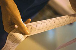
An EKG or ECG reveals how your heart's electrical system is working. The ECG senses and records your heartbeats, or heart rhythms. The results are printed on a strip of paper. An ECG can also help your doctor diagnose whether:
- You have arrhythmias
- Your heart medication is effective
- Blocked coronary arteries in the heart are cutting off blood and oxygen to your heart muscle
- Your blocked coronary arteries have caused a heart attack.
In all, there are three kinds of tests that record your heart's electrical activity, each for a different period of time:
- Electrocardiogram (EKG/ECG) done in the doctor's office. It records your heart rhythms for a few minutes.
- Holter monitoring - records and stores (in its memory) all of your heart rhythms for 24 to 48 hours.
- Event recorder - constantly records your heart rhythms. In contrast to a holter monitor, it stores the rhythms (in its memory) only when you push a button.
What are the parts of an ECG Strip?
The peaks on an electrocardiogram (ECG) strip are called waves. Together, all the peaks and valleys give your doctor important information about how your heart is working. The P-wave shows your heart's upper chambers (atria) contracting. The QRS complex shows your heart's lower chambers (ventricles) contracting. The T-wave shows your heart's ventricles relaxing.
How is an ECG Performed?
When you have an electrocardiogram (ECG) you undress from the waist up, put on a hospital gown, and lie on an exam table. As many as 12 small patches called electrodes are placed on your chest, neck, arms, and legs. The electrodes, which connect to wires on the ECG machine, sense the heart's electrical signals. The machine then traces your heart's rhythm on a strip of graph paper.